Microscopes can see what is invisible to the naked eye. For this reason, and many others, they are essential for scientists who aim the learn more about the way the human body operates by studying those minuscule building blocks called cells.
A single sample of saliva or blood, containing millions of cells, can reveal a lot about a person’s health conditions. A particular blood test called the Peripheral Blood Smear review, for example, the test is conducted by a human looking at a sample under a microscope, methodically classifying cells by type, counting them, and checking for anomalies that could even mean cancer. Pen and paper at hand ready to take notes. Jotting down numbers. A stiffening, monotonous and tiring procedure.
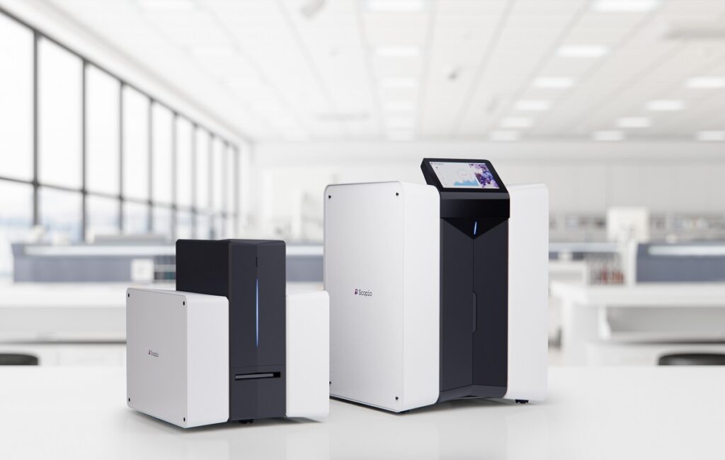
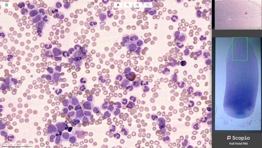
This image, so emblematic of lab work, is on its way to becoming a thing of the past. Scopio Labs, an Israeli company founded in 2015, has developed a digital imaging platform that harnesses computational photography to capture the full field at 100X resolution, and an AI-powered Decision Support System (DSS) to digitize, quantify, and analyze hematology and cytology cell samples with ultra-high resolution, thereby making the identification of potential cell abnormalities easier and more precise without all the headache and work.

Scopio Labs’ CTO Erez Naaman, a 39-year-old graduate of the elite IDF R&D unit Talpiot, tells NoCamels over Zoom how the company was co-founded with pal and Scopio Labs CEO Itai Hayut. They thought about ways to streamline the common blood test for pathologists.
“It’s the most common test in the world, it’s done in every lab. And blood flows through all of the different organs in your body. It interacts and reacts with everything. So when you look at the morphology [of blood cells], you’re actually able to see what’s happening in your body. Right now. You can see the cells are reacting to a virus, to a bacterium, to cancer. There are many different things that happen in the blood,” says Naaman.
The reality the two friends were faced with on their first trip to hospital left them astounded. “We entered a room and we [saw] 20 people sitting over microscopes, counting cells and writing manually, not even on a computer, with pen or pencil on a piece of paper. And we thought that this doesn’t make sense!” he explains.
The Tel Aviv-based company, which was founded in 2015, just raised a $50 million Series C round in February to further develop the diagnostic imaging tool. The company has already received FDA clearance and a CE mark for its X100 Microscope and Decision Support System With Full Field Peripheral Blood Smear (PBS) Application, making it possible to use the platform in laboratories across the US and Europe.
Lego and Blu-ray
Blood analysis is laborious because conventional microscopes allow no compromise between a large, low-resolution and a narrow but high-resolution field of view. The clinician can therefore only focus on one parcel of the blood sample at a time. Furthermore, microscopes cannot capture pictures of the magnification for later cross-checking. The clinician has to manually count and detect anomalies in the blood on the spot by looking through the microscope’s lenses.
Unable to shake off the image from the hospital, Naaman and Hayut began wondering whether one could develop a scanning microscope with an AI feature that could do the counting of cells instead.
“We took over Itai’s apartment and made it into a lab. We began building a scanning microscope. We hacked into Blu- ray devices and started assembling something with Lego and 3D-printed parts. And as we were doing it, we realized why there was no scanning microscope at very high resolution. And that was because the mechanics and optics required are just really difficult,” he admits.
Sign up for our free weekly newsletter
SubscribeInstead, they decided to use computational photography to fully capture the details of broad low-resolution scans. “Computational photography is the combination of hardware and software to bypass the classical limitations of the hardware. Before us, the problem was, how do you get the mechanics and the optics to work. We turned it into a software problem,” Naaman says.
“Using software to enhance images may sound very high-tech, but most people are already exposed to basic versions of hardware-enhancing technology through their smartphones,” says Naaman, “In fact, high-resolution selfies, landscape views and group shots would be inconceivable without the underlying digital software that supports the work of our tiny smartphone lenses.“
“The trick here is to understand how light behaves. Essentially, what we do is we take a lens that has a large field of view and low resolution. We take multiple images of that same area, under different illumination conditions, and at different angles, each one designed to give us a little more information about the sample. And then what we do is we actually have a physics-based model that takes those images and reconstructs the entire field of light, including the invisible parts of the light. Once we know the actual field of light, we can perform super-resolution on the [sample scan] from all those images,” he adds.
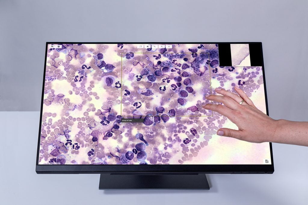
The result is a printer-like machine capable of providing lab physicians with a full-field high-resolution picture of the blood sample to be displayed on their computers or tablets. In one scanned frame, the clinician can zoom in and out to the very fringes of the sample without missing any details.
Naaman recalls the initial setbacks faced when developing their first prototype. “It wasn’t stable. It was so sensitive that every time we wanted to run it, we had to [do a countdown] and hold our breath. And if anyone accidentally moved, then we had to restart it. It was also very slow. The first working version would have taken three years to compute the information from one sample. Today, we do a sample in minutes.”
Finding Waldo or the future of AI in medicine
Scopio Labs’ technology does more than just scan blood samples. “Our technology is not just a digital microscope, it’s a diagnostic platform. Scanning and creating the digital file has merit. But the combination of the data with the layer of analysis on top of the data is where the real magic lies,” says Naaman.
The scan provided by the imaging platform is analyzed by AI, which can count, organize and arrange cell types and inform of anomalies in the blood sample. “Our decision support system can pre-analyze the sample and provide the results to the [lab employees] both locally and remotely. Our focus is to basically take out all of the tedious parts of the operation. You can’t tell a person: ‘Count 150,000 of these cells and tell me if two of them are different.’ It’s an impractical task. It’s like ‘Where’s Waldo?’ Looking for something hidden in data is really difficult for a person. For a computer that has been taught, it takes ten milliseconds. Every time, no matter what, it’ll always find Waldo,” says Naaman.

Yet, while Scopio Labs aims to replace the use of the manual microscopes for cell analysis, it does not want to replace the people sitting behind it. “Our aim is to enhance the capability of the clinicians and the lab employees by taking away the tedious parts of their work and provide them with all the information that they need to enable early detection and monitoring of medical conditions that manifest themselves in blood. People are really good at drawing conclusions, way better than computers. We want to leave the people in charge of combining the complicated circumstances into a clinical decision. But the task of finding cells, classifying them, organizing them, that’s a task that computers do better than people. So we don’t take away the clinical aspect, we actually empower it,” he adds.
When asked about the future of AI in medicine, Naaman answered that it will change how medical staff will work in the future. “The efficiency of each person in the medical world will be enhanced tenfold and more with what AI can do, because these people won’t waste their time [collecting] the data. They’ll spend their time using the data. When we get to a critical mass of enough [end-to-end] applications where you don’t have to go into separate systems to do different [health checks], or you do only this with AI and the other things are not approved, I think that’s when the revolution [in medicine] will happen.”
Related posts

Israeli Medical Technologies That Could Change The World

Harnessing Our Own Bodies For Side Effect-Free Weight Loss

Missing Protein Could Unlock Treatment For Aggressive Lung Cancer


Facebook comments