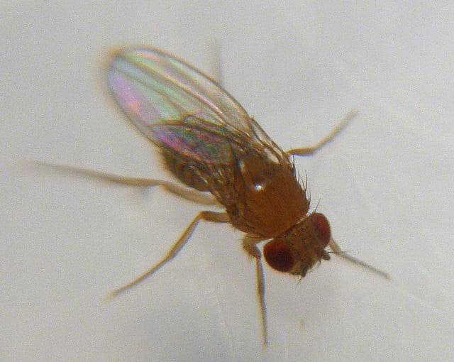
Imagine a human brain being kept alive in a vat for research. That vision might be for a distant future, but researchers at Tel Aviv University have pioneered a technique for developing neurons, the building blocks of the brain, in petri-dishes. The neurons are from flies know as fruit fly and the technique is being used to study Alzheimer’s disease.
Alzheimer’s may be a well-known disease worldwide, but it is difficult to study in humans. A little-known fact is that positive confirmation of Alzheimer’s disease in humans can only occur through a brain autopsy after the person has died, greatly impeding research on the disease.
Related articles
- New Application Allows You To Create Life On your Computer
- Haifa University Researchers Find Important Link In Fight Against Alzheimer’s
“As far as we know, the neuron, as a building block of the brain, is the same whether it is in a human or a cockroach,” says Professor Amir Ayali at Tel Aviv University’s Department of Zoology and Sagol School of Neurosciences. He tells NoCamels: “The insect brain, being simpler, is thus appropriate for examining how neurons develop at the basic level.”
“Growing” a two-dimensional brain
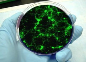
What makes the technique special is the ability to observe neural development at the cellular level. In the lab, the researchers break the fly’s nervous system down into single cells, separate these cells, and then place them at a distance from each other in a Petri dish.
After a few days, the neurons begin to grow towards one another and establish connections, and then migrate to form clusters of cells. Finally, they re-organize themselves to form a sophisticated network. The two-dimensional neural culture allows researchers to concentrate on individual neurons, and they can perform specific measurements of proteins, note electrical activity, watch synapses develop, and see how physical changes take shape.
To apply the technique to the study of Alzheimer’s disease, researchers compare the measurements taken from the neuron development of a normal fruit-fly and the measurements taken from the neuron development of a special strain of fruit-fly. This special strain of fruit-fly has been genetically modified to show symptoms of Alzheimer’s.
Sign up for our free weekly newsletter
SubscribeThe fly is modified so that it expresses a peptide called Amyloid Beta, found in protein-based plaques of humans with Alzheimer’s disease. The genetically-modified fruit-flies demonstrate Alzheimer’s-like symptoms such as motor problems, impaired learning capabilities, and shorter lifespans.
According to Ya’ara Saad, a PhD candidate working in Ayali’s lab, researchers will compare the structure, the protein make-up and the activity of the two cultured neural networks in order to find out how Amyloid Beta interferes in neural development.
Testing drugs on neural cultures
The use of neuron culture extends beyond researching on how Amyloid Beta affects the brain. Potential drugs that fight Alzheimer’s by inhibiting Amyloid Beta can also be tested for effectiveness using the neural cultures. In addition, other neurological disorders can be studied in a similar way to that of Alzheimer’s. Generally, all that is needed is to change the strain of fly that is used to one that expresses the neurological disorder to be studied.
“It all began with the simple question of ‘how do neurons develop?’” says Ayali. “To answer that question, we started developing neural cultures of the locust. Then, we decided to use that technique on fruit flies because there has been so much research done on them.” Obviously, this is a technique that holds potential for further research and discovery.
Photo by Wanderin’ Weeta
Related posts

Israeli Medical Technologies That Could Change The World

Harnessing Our Own Bodies For Side Effect-Free Weight Loss

Missing Protein Could Unlock Treatment For Aggressive Lung Cancer


Facebook comments