For thousands of years, microscopes have allowed scientists, doctors, and lab professionals to view enlarged images of small objects, cells, bacteria, and other agents that cannot be seen by the naked eye, for the purpose of analysis and examination. Since the invention of the first microscope around 1590, these devices have come a long way, allowing for varying levels of magnifying power and producing images of different types and resolutions. The “world’s most advanced microscope” is said to be able to probe the spaces between atoms.
But microscopes – specifically modern research ones called compound microscopes – remain largely analog devices.
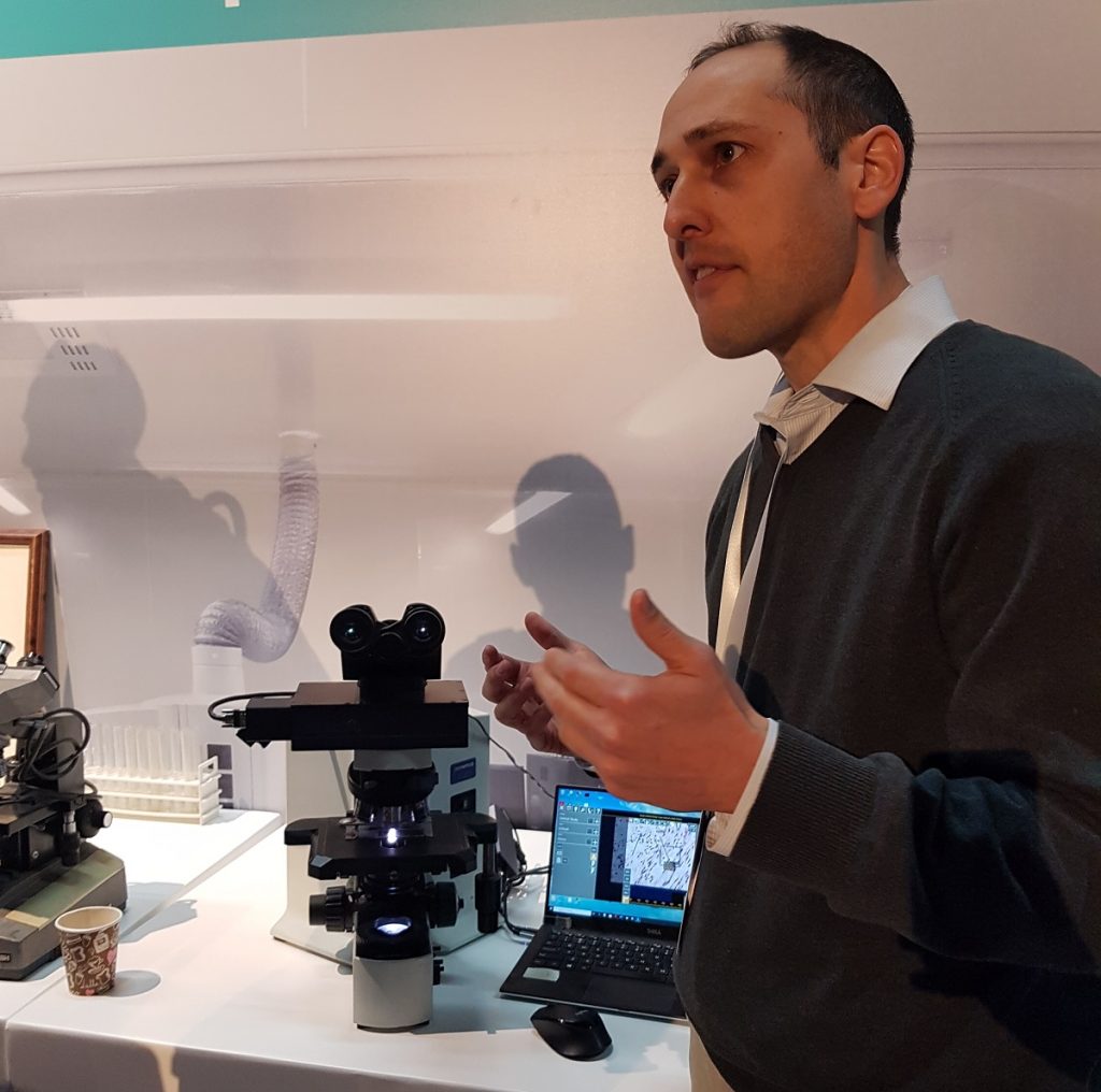
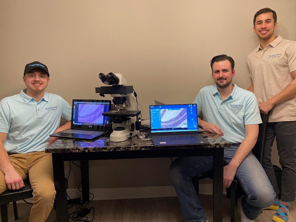
Augmentiqs, an Israeli startup founded in 2016 and based in the northern city of Misgav, seeks to bring microscopes into the modern millennium by turning these medical instruments into augmented reality (AR) devices that will allow for smart, real-time digital pathology.
Pathology is the branch of medical science that involves the study and diagnosis of disease through the examination of certain aspects of the body. The microscope is still the instrument of choice for pathologists.
For digital pathology, a sub-field of pathology that focuses on data management based on information generated from digitized specimen slides, virtual microscopy – posting microscope image on, and transmitting them over, computer networks – is the preferred method of examination.
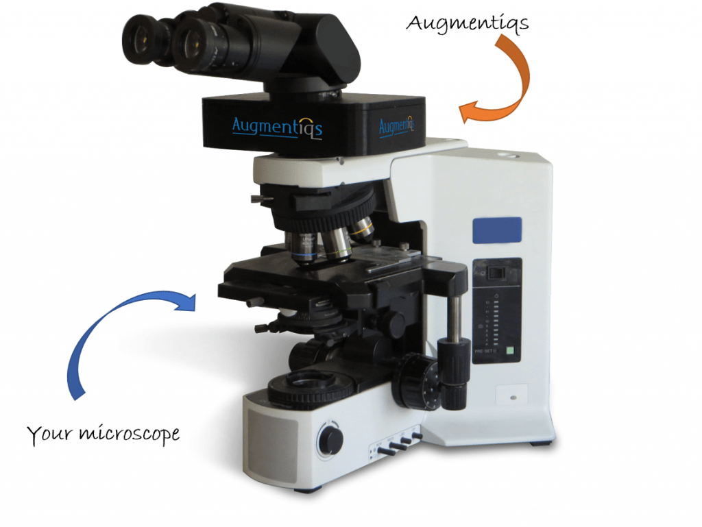
More specifically, Augmentiqs’ digital pathology solution embraces and enhances the existing microscope, helping pathologists work more effectively, connect remotely with colleagues, and conduct groundbreaking research, the company says.

Augmentiqs’ solution bridges the gap between the analog microscope and the digital world, the company’s CEO and co-founder Gabe Siegel tells NoCamels.
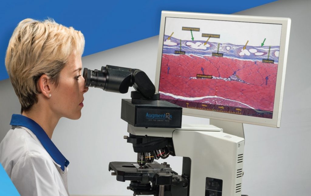
Augmentiqs’ system is integrated into an existing microscope, connecting it to a PC and transforming it into a smart interactive device that offers a cost-effective alternative to digital pathology, according to the company. This enables real-time examination through the augmented projection of what the microscope eyepiece actually sees.
For pathologists, the platform drastically improves what microscopes have to offer to generate a pathology that is cheaper, faster, and more accurate.
“We came up with a low-cost solution for a digital microscope that doesn’t change the microscope, but enhances it, by providing capabilities of a computer straight to the microscope,” Siegel explains.
From traditional tech to ‘smart’ tech
Siegel says it was the level of distrust he experienced from pathologists at trade shows while working for a different digital pathology company that made him realize digital pathology tech needed to be improved.

“A pathologist comes over and says, ‘In 90 percent of instances, I don’t need you. And in 10 percent, I don’t trust you.’ It was experiences like these that made us realize that the digital pathology tech out there doesn’t fit the needs of the pathologists,” Siegel tells NoCamels.
Augmentiqs sought to revolutionize the digital pathology industry.
Sign up for our free weekly newsletter
Subscribe
The tech in the Augmentiqs system enables pathologists with the ability to interact with the specimen being examined while viewing it at the same time, Siegel explains. They can also choose which software to run according to their needs. The digital software acts as a platform where pathologists can measure, annotate, and count cells. It can also run AI algorithms.
The system gives pathologists unlimited applications for improved clinical data collections using the inbuilt software QuPath. This and many other applications can be used as tools to help a pathologist detect specific cells, such as cancer cells and accurately count how many are present.
“We are very adaptive to the real needs of the pathologists,” Siegel says. “The idea is how do we provide clinical value that fits the workflow and also not be cost-prohibitive.”
According to Siegel, each pathologist “is going to find a different functionality based on what they need to accomplish and this is the reason why they would want to incorporate this technology,” he explains. It gives them a wide range of options that fit the unique requirements of different pathologists, he adds.
The Augmentiqs system uses high-resolution cameras in order to generate augmentations that can be shared and viewed by multiple pathologists at the same time. This provides labs worldwide with the ability for a real-time telepathology (sharing of pathological data through telecommunications).
Augmentiqs also tracks which coordinates of the slide are being examined, which allows other viewers to reexamine the same slide areas that were initially analyzed. It does this by projecting directions within the eyepiece that can be followed by pathologists. (Siegel pointed out that the tech could also serve in microscopy education so students can follow along.)
In addition, medical researchers and AI developers can also create their own applications for the pathology ecosystem using Augmentiqs’ Open API system.
Another feature, which Siegel says is “totally new,” gives clinical informatics professionals, developers and other pathology software providers the ability to create and deploy their own algorithms directly within the microscope. In September, Augmentiqs entered into a partnership with Aira Matrix, an American company that provides image analysis and management solutions for preclinical toxicology and pathology applications, to complete the first preclinical pathology study that utilizes AI algorithms in the microscopic examination.
Siegel admits that in order for the company to grow, educating the market on the importance of Augmentiqs digital software and tech to improve pathology results is key. The company is currently in the seed funding stage.
SEE ALSO: Technion Researchers Improve Microscope Resolution Tenfold
“Our method of education is focused more on trying to get studies and scientific journals published by the users that have already incorporated the product into their workflow. When we have the pathologists using our product, we enable them to generate studies that haven’t been achieved before,” Siegel says.
Meanwhile, the reception from pathologists who use the platform has been promising. “Every pathologist who’s seen it has been totally amazed; the feedback from them has always been absolutely positive,” Siegel tells NoCamels.
Related posts

Editors’ & Readers’ Choice: 10 Favorite NoCamels Articles

Forward Facing: What Does The Future Hold For Israeli High-Tech?

Impact Innovation: Israeli Startups That Could Shape Our Future


Facebook comments