Israeli researchers have developed a way to model human muscle diseases in tiny worms, providing a new, versatile platform to study such ailments.
The researchers from Bar-Ilan University and Sheba Medical Center took extracellular vesicles (very small sacs of fluid released by cells to perform a range of functions) from the blood of patients with Duchenne muscular dystrophy.
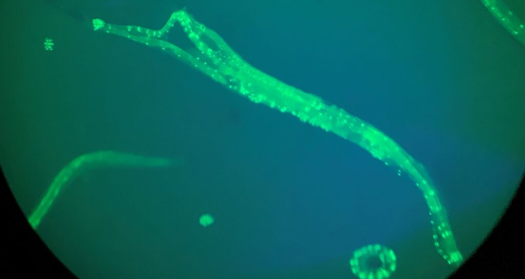
When the vesicles were transferred to C. elegans worms, it led to muscular atrophy closely resembling human symptoms of the disease.
“Unlike traditional methods that rely on genetic modifications in model organisms, this technique relies on elements secreted into the blood,” said Bar-Ilan doctoral student Rewayd Shalash, who carried out the research with fellow doctoral student Coral Cohen and Dr. Mor Levi-Ferber from the university’s Goodman Faculty of Life Sciences.
The study was led by Prof. Chaya Brodie and Prof. Sivan Korenblit, both from the Goodman Faculty, alongside Dr. Amir Dori, a muscular dystrophy specialist from Sheba, the largest medical center both in Israel and the entire Middle East.
“We transferred the extracellular vesicles and observed the development of muscular dystrophy with significant muscle degeneration, confirming that we discovered a new way to model diseases without the need to alter specific genes,” said Korenblit.
Related posts

Israeli AI Safety Tool Among TIME’S Best Inventions For 2024

TAU Team Discovers Mechanism To Eliminate Cancerous Tumors

Ashdod Port Investing In Startups As Part Of Innovation Strategy


Facebook comments