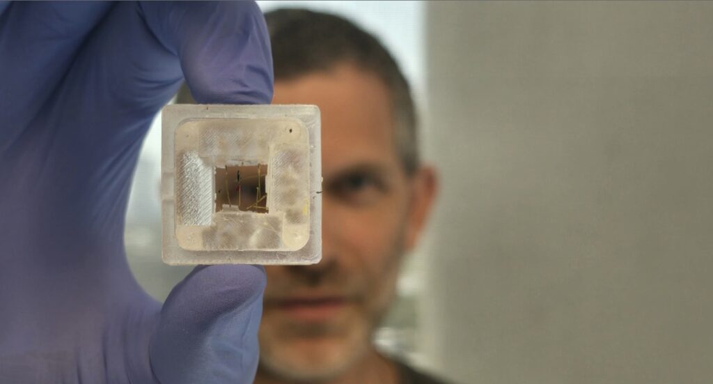
Researchers at Tel Aviv University have used the principles of origami, the Japanese art of paperfolding, to develop a solution for the problem of how to accurately place sensors inside models comprising 3D-printed tissue.
The solution helps scientists resolve the long-standing problem of being unable to bioprint the tissue over the sensors, which are used to provide information from inside the model for research purposes.
Using an origami-inspired structure that folds around the printed tissue, the scientists found that they could insert the sensors into exact defined locations in order to monitor electrical activity or resistance of cells.
“In this study, we created an ‘out-of-the-box’ synergy between scientific research and art,” they said.
And while demonstrations of the new system, dubbed the Multi-Sensor Origami Platform, focused on brain tissue, the researchers say that it can place any number and type of sensors in any location within any form of 3D-bioprinted tissue model.
The research was a joint effort by members of several of the university’s departments, including the School of Neurobiology, Biochemistry and Biophysics; Koum Center for Nanoscience and Nanotechnology; and Department of Biomedical Engineering.
“The use of 3D-bioprinters to print biological tissue models for research is already widespread,” explained Prof. Ben Maoz of the Department of Biomedical Engineering, who was one of the researchers.
“In existing technologies, the printer head moves back and forth, printing layer upon layer of the required tissue. This method, however, has a significant drawback: The tissue cannot be bio-printed over a set of sensors needed to provide information about its inner cells, because in the process of printing the printer head breaks the sensors. We propose a new approach to the complex problem: origami,” he said.
Related posts

Israeli AI Safety Tool Among TIME’S Best Inventions For 2024

TAU Team Discovers Mechanism To Eliminate Cancerous Tumors

Ashdod Port Investing In Startups As Part Of Innovation Strategy


Facebook comments