For decades, doctors and surgeons have been relying on medical imaging to help make diagnostic decisions, guide medical interventions, and decide on courses of treatment. Techniques such as positron emission tomography (PET) and X-ray radiology applications like CT scans and MRIs are often used to diagnose and treat diseases as well as analyze the body’s internal structures.
The field of holography, the science and practice of making holograms, is relatively new to the medical world but it holds much promise. And Israeli company RealView Imaging is looking to create a new dimension for medical imaging applications with its pioneering hologram system for the human body.
SEE ALSO: Israeli Firm’s Holographic Imaging Tech Used In 1st Live Medical Procedure
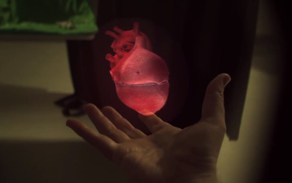
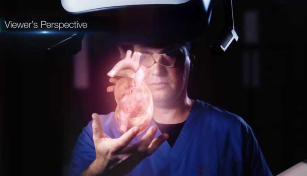
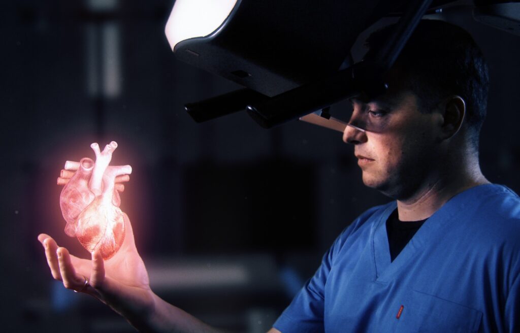
First founded in 2008 by Aviad Kaufman, Shaul Gelman, and Professor Carmel Rotschild, RealView developed the Holoscope-i, the world’s first medical holographic system that provides spatially accurate 3D in-air holograms of organs. The system is designed for physicians to view and hyper-realistic 3D holograms of the patient’s actual anatomy during interventional procedures.
“We’re the only system in the world that was developed to provide augmented reality only for physicians for very very specific use cases…” Kaufman tells NoCamels. “It is a very accurate and realistic image by taking the anatomy of the patient out of the patient and showing it in the air.”
“[Physicians] also have the ability to interact with this information because when you have a hologram in front of you, you can now take your finger inside, you can cut it, you can mark it, you can manipulate the image,” Kaufman adds.
In 2013, RealView partnered with Dutch multinational Philips to demonstrate the feasibility of using its live 3D holographic visualization and interaction system to guide minimally-invasive structural heart disease procedures. The collaboration was part of a clinical study at the Schneider Children’s Medical Center in Petach Tikva where doctors were able o view detailed dynamic 3D holographic images of the heart and manipulate the structures by “literally touching the holographic volumes in front of them.”
Dr. Elchanan Bruckheimer, pediatric cardiologist and Director of the Cardiac Catheterization Laboratories at Schneider Children’s Medical Center, said at the time: “The ability to reach into the image and apply markings on the soft tissue anatomy in the X-ray and 3D ultrasound images would be extremely useful for guidance of these complex procedures.”
A 2016 study by Dr. Bruckheimer and the RealView founders successfully demonstrated the technology for the first time in clinical medical imaging.

Three years later, the Holoscope-i was used in the first live medical procedure at the Toronto General Hospital’s Peter Munk Cardiac Centre (PMCC) where cardiologists and cardiac surgeons performed a valve-in-valve mitral valve procedure, a minimally invasive procedure that replaced a worn-out surgical valve. Israeli President Reuven Rivlin attended the Toronto unveiling, and PMCC noted that it is using the hologram for other cardiac procedures, such as repairing leaking valves and closing holes in the heart.
A growing market for medical holography
RealView Imaging is collaborating with the largest medical imaging companies to provide them with this revolutionary technology, Kaufman indicates. The global digital holography market is projected to reach $5.4 billion by 2024 from $2.2 billion in 2019, according to a Markets and Markets report released this year.
Kaufman says holography is the best method in science to precisely reconstruct and display 3D objects in air. Since holograms are not optical illusions but rather optical realities, it is very difficult to distinguish between a high-quality reconstructed hologram and the original object.
According to RealView, current augmented reality solutions often inaccurately consider themselves holograms without actually using real, inference-based holography. Additionally, current solutions allow users to see and interact with 3D images from a distance without being able to manipulate them and sometimes give users an actual headache due the vergence-accommodation conflict, which occurs when the brain receives mismatching cues between the distance of a virtual 3D object and the focusing distance.
Sign up for our free weekly newsletter
Subscribe“Go to any expert in 3D imaging and [ask] what is the best way to reconstruct a 3D image. The answer will undoubtedly be a real volumetric interference-based hologram, so there’s no debate in the scientific world that this is the best method to reconstruct an image, but they will also tell you that it’s very hard to complete it, it’s almost impossible to do it in real time with high quality,” Kaufman tells NoCamels.
The Holoscope-i can be used for long periods of time “which physicians need when they’re doing complicated procedures, and with no headache or fatigue at all or nausea, because I’m not pulling the brain in any way. We’re really providing them with an issue that is very accurate,” Kaufman explains.
Holograms and more
RealView’s proprietary Digital Light Shaping technology creates a unique hyper-realistic experience for visualization of medical images, specifically through the Holoscope-i, that provides full color, high resolution, dynamic, and interactive 3D images in free space from any medical 3D volumetric data. The system has been ergonomically designed for use in all clinical environments like interventional suites, hybrid operating rooms and diagnostic clinics.
It allows physicians to visualize the fully volumetric 3D hologram to intuitively understand each organ’s anatomy. Physicians can also rotate, slice, mark, and measure the holograms to examine internal anatomy to scale. The holograms are projected from overhead systems mounted above the physician, allowing them to better understand the patient’s complex spatial anatomy and dynamic physiological processes for more effective treatment and outcome.

RealView already has 21 registered patents with another 14 pending. Its tech has already been demonstrated at international trade shows, and Kaufman hopes to have multiple clinical trials in the United States within the next year.
Last month, RealView raised $10 million in Series C funding and announced the development of a new system, the Holoscope-x, which aims to display the hologram within the patient’s body, making the patient literally “see-through.”
Kaufman notes that Holoscope-x is of great clinical value especially for interventional radiology procedures that rely on 3D imaging. It will allow physicians to improve the accuracy and efficacy of procedures and better understand spatial anatomy and collateral vasculature.
SEE ALSO: Israeli Medical Holography Firm Raises $10M For 3D in-Air Hologram System
“Imagine you have a patient that has a tumor, and you need to do a biopsy procedure… Today, the tumor is 3D, and we have the specific information of the tumor, and what we’re able to do because we’re projecting the hologram to your eye is to project the tumor and the tools that are targeting the tumor inside the patient,” Kaufman says. “Imagine that you can look into a patient and see the tumor inside, the organs inside, the bones, etc.”
Kaufman tells NoCamels that this technology can potentially save many lives since “many times physicians spend a lot of time navigating” the patient’s organs without a clear visualization of their anatomy. In critical operations, physicians can, in real-time, locate exactly where they must operate much more independently to save a patient’s life.
“Better imaging provides, at the end of the day, better clinical results and this is the path that we are working to continue to prove,” Kaufman says.
Related posts

Editors’ & Readers’ Choice: 10 Favorite NoCamels Articles

Forward Facing: What Does The Future Hold For Israeli High-Tech?

Impact Innovation: Israeli Startups That Could Shape Our Future


Facebook comments